Mumbai
08048056620
+919833066702
about-cover-bg image
About Dr. Nihar Modi

The Best Orthopaedic Doctor & Bone Specialist in Mumbai

Treatments
Featured Treatments

What We Cure

Fracture Fixation

Fracture fixation is a surgical method used to stabilise a broken bone when non-operative treatment is unlikely to provide adequate alignment, stability, or healing. Fractures may occur because of falls, sports injuries, road traffic accidents, direct trauma, or weakened bone. Symptoms commonly include sudden pain, swelling, bruising, tenderness, difficulty using the limb, and sometimes visible deformity. The need for fixation depends on the type and location of fracture rather than pain alone. Evaluation usually includes a physical examination and imaging. X-rays are the primary investigation for many fractures, while CT may be used for complex fractures or injuries involving a joint. The clinician also checks circulation, sensation, and movement because fractures can be associated with nerve or blood-vessel injury. Open fractures require urgent specialist treatment because the broken bone or wound may communicate with the outside environment and carry an increased risk of infection. Fracture fixation can use plates and screws, intramedullary rods or nails, wires, pins, or other devices depending on the bone and fracture pattern. The aim is to restore appropriate alignment and provide enough stability for the bone to heal while allowing rehabilitation to begin at a suitable stage. Not every fracture requires surgery; stable injuries may be managed with a cast, splint, or brace. After fixation, care may involve wound monitoring, pain management, protection of the repaired area, and progressive physiotherapy. Weight-bearing or loading restrictions vary according to the fracture, fixation method, bone quality, and healing progress. Follow-up appointments and imaging may be used to monitor alignment and bone healing. Rehabilitation then focuses on restoring range of motion, strength, balance, and functional use. Patients should seek urgent medical attention after an injury if there is an obvious deformity, severe uncontrolled pain, an open wound over a suspected fracture, loss of sensation or movement, or a cold, pale, or blue extremity. These signs may indicate serious injury requiring immediate care. This information is for general education and is not a diagnosis or personalised treatment plan. Consult a qualified orthopaedic doctor before choosing medical care. Seek emergency care for severe or potentially life-threatening symptoms.

Tendon Repair

Tendons connect muscles to bones and transmit the force needed for movement. Tendon injuries can occur suddenly, such as after a sports injury or fall, or develop gradually through repetitive loading and degeneration. Depending on the tendon involved, symptoms may include pain, swelling, weakness, tenderness, reduced movement, difficulty using the affected limb, or a sudden loss of function after an acute tear. A complete tendon rupture can produce a more pronounced functional deficit than a minor or partial injury. Diagnosis usually begins with a history and physical examination. The clinician assesses the mechanism of injury, strength, range of motion, and tendon continuity. Ultrasound or MRI may be used when the diagnosis is uncertain or when the extent of injury needs to be defined. Treatment depends on the tendon, location and severity of the injury, whether the tear is partial or complete, the patient's functional requirements, and other health factors. Some tendon problems can improve with activity modification, physiotherapy, progressive loading, and appropriate pain management. Rehabilitation aims to restore tendon capacity, strength, movement, and function. Selected complete tears or injuries associated with substantial functional loss may require surgical repair. During tendon repair, the damaged tendon ends are brought together using appropriate surgical techniques. The repaired tendon then requires protection while biological healing occurs. Postoperative rehabilitation is usually gradual. Early treatment may prioritise protecting the repair and maintaining safe movement. Later stages progressively restore strength and function under professional guidance. Returning too quickly to heavy loading or sport can place excessive stress on a healing tendon, so activity progression should follow the treating team's protocol. This information is for general education and is not a diagnosis or personalised treatment plan. Consult a qualified orthopaedic doctor before choosing medical care. Seek emergency care for severe or potentially life-threatening symptoms. Prompt assessment is recommended after an acute injury that causes sudden loss of function, a noticeable gap or deformity, severe pain, or significant swelling. Numbness, weakness, or signs of impaired circulation require urgent evaluation. The rehabilitation plan is adjusted to the tendon involved because different tendons have different healing and loading requirements.

Cartilage Restoration

Cartilage restoration refers to procedures and treatment strategies intended to address focal damage to articular cartilage, the smooth tissue that helps joint surfaces move with low friction. Cartilage injuries can occur after trauma, sports injuries, repetitive loading, or other joint problems. Symptoms may include activity-related pain, swelling, stiffness, catching, reduced range of motion, or difficulty participating in sport. Cartilage damage can coexist with ligament or meniscal injuries, so a complete assessment is important. Evaluation generally includes a clinical history and physical examination followed by appropriate imaging. X-rays can help assess alignment and arthritis, while MRI may provide information about cartilage, bone, ligaments, and other soft tissues. Treatment depends on the location, size, depth, and pattern of cartilage damage, the patient's age and activity level, joint alignment, and whether other structures are injured. Non-operative care may include activity modification, physiotherapy, strengthening, weight management when appropriate, and pain management. Some focal cartilage defects may be considered for joint-preserving procedures when symptoms persist and the overall joint is suitable. Depending on the defect and clinical circumstances, surgical approaches can include techniques that stimulate repair or procedures that restore or replace damaged cartilage tissue. These are different from joint replacement and are intended to preserve the patient's joint in selected situations. Recovery depends heavily on the procedure performed and the location of the cartilage defect. Some treatments require a period of protected weight-bearing or restrictions on high-impact activity so that the repaired area can heal. Rehabilitation may progress through controlled movement, muscle strengthening, balance, and gradual return to functional or sporting activity. Patients should follow the specific rehabilitation protocol provided by their treating team rather than assuming that all cartilage procedures have the same recovery schedule. Cartilage restoration is not appropriate for every patient or every form of arthritis. Advanced degenerative joint disease may require a different treatment strategy. A specialist assessment is therefore important before selecting a procedure. This information is for general education and is not a diagnosis or personalised treatment plan. Consult a qualified orthopaedic doctor before choosing medical care. Seek emergency care for severe or potentially life-threatening symptoms.

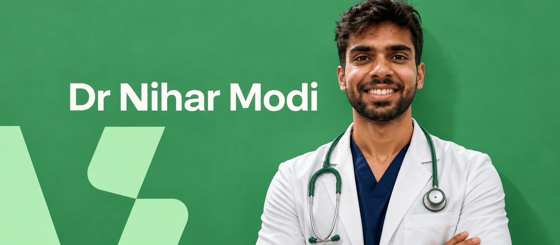
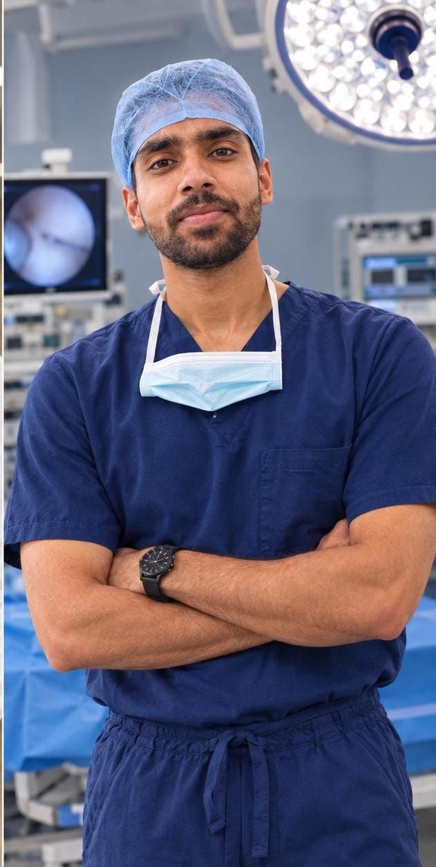
Doctor's Profile Picture

Dr Nihar Modi

Dr Nihar Modi

M. S. Ortho | D.N.B. Ortho | MRCS (England) | Diploma in Football Medicine (FIFA) | Fellowship in Shoulder, Elbow & Knee Sports injuries, Arthroscopy and Arthroplasty (Australia, USA)
Mumbai

Speciality

Orthopaedic Surgeon


Education

M. S. Ortho | D.N.B. Ortho | MRCS (England) | Diploma in Football Medicine (FIFA) | Fellowship in Shoulder, Elbow & Knee Sports injuries, Arthroscopy and Arthroplasty (Australia, USA)


Experience

12 years


Mobile

9833066702


Memberships

NA


Registration No

9833066702

Image Gallery

Pictures That Speak

Need Trusted Second Opinion?

Not sure about your diagnosis or treatment plan? We're here to help.

Testimonials View All
testimonial image

Severe knee instability had made it difficult for me to even walk down the stairs without fear. Dr. Nihar Modi diagnosed...

Read More
testimonial image

The doctor is incredibly polite and compassionate. Treatment is concise and ethical. Highly recommended. Thank you, Dr N...

Read More
testimonial image

Doctor is very knowledgeable. I had a sprain in my ankle when i visited him, helped me step by step!

footerhc